
Abstract
Introduction: Breast carcinoma is the second most
common malignant tumor and leading cause of
death in women. The fine needle aspiration cytology of breast lump is a rapid, safe, cost effective and good screening method .It is important tool for
initial diagnosis and management of palpable breast
swellings. The aim of this study is to identify the
common benign and malignant tumors of the breast
lump.
Materials and methods: A retrospective longitudinal study was carried out in the Department of Pathology, Pokhara Academy of Health Sciences
over one year period. Procedure was done using 10
ml disposable syringe. The air-dried smear and wet
smear was stained with Giemsa stain, papanicolaou
stain respectively for morphological study.
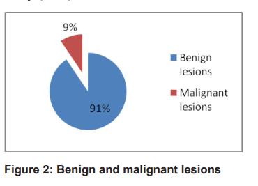
Results: Out of 95 cases, 93 were females. Breast
lumps were most common in 20 to 40 years of age
group i.e. 48.42 %( n=95). Benign lesions were
found in 91%. In 22.10% (n=95) of cases, fibroadenoma was seen followed by benign chronic inflammatory lesion (18.94%). In malignant tumor, ductal
carcinoma was diagnosed mostly in 40 -80 years
age group.
Conclusion: Our study concluded that fine needle
aspiration cytology is the early diagnostic tool for
diagnosis of benign and malignant tumor. Fibroadenoma was the common benign tumor and invasive ductal carcinoma was the common malignant tumor.
References
Binu, V. S. et al. Cancerpatterns nepal.Pdf.
, 183–186 (2007).
WRC. Table of Contents Table of Contents
.Univ. Eur. ا ز س ی ر ت ا پ ی ا ز م ص ا ح ب ه د ک ت ر ی
Inst. 2–5 (2012).
Shrestha, A., Chalise, S., Karki, S. &
Shakya, G. Fine needle aspiration cytology
in a palpable breast lesion. J. Pathol. Nepal1,
–135 (1970).
Choudhary, P., Koirala, A., Rimal, H. & Deo,
A. Cytomorphological study of palpable
breast lumps. J. Pathol. Nepal5, 817–819
(2015).
Rathod, G. B., Jain, M., Vachhani, D. &
Balar, M. Retrospective study of fine needle
aspiration cytology of clinically palpable
breast lump. 3, 69–73 (2016).
Bukhari, M. H. et al. Use of Fine-Needle
Aspiration in the Evaluation of Breast
Lumps. 2011, (2011).
Sharif, A., Tabassum, T., Riaz, M., Akram,
M. & Munir, N. Cytomorphological patterns
of palpable breast lesions diagnosed on fine
needle aspiration cytology in females. Eur.
J. Inflamm.18, (2020).
~448~˷
Original Article Medical Journal of Pokhara Academy of Health Sciences Vol 5 Issue 1 Jan-Jun 2022
Badge, S., Ovhal, A., Azad, K. & Meshram,
A. Study of fine-needle aspiration cytology
of lymph node in rural area of Bastar
District, Chhattisgarh. Med. J. Dr. D.Y. Patil
Univ.10, 143–148 (2017).
Pathak, S., Saxena, A. K., Khan, S. & Jha,
J. K. Cytomorphological Study of Palpable
Breast Lumps by FNAC. Int. J. Contemp.
Pathol.4, 16 (2018).
Article, O. Study of Fine-Needle Aspiration
Cytology of Breast Lumps in Rural Area
of Bastar District, Chhattisgarh. 339–
(2017) doi:10.4103/MJDRDYPU.
MJDRDYPU.
Gupta, R., Dewan, D., Kumar, D. & Sharma,
R. Utility of fine-needle aspiration cytology
as a screening tool in diagnosis of breast
lumps. Int. Surg. J.4, 1171 (2017).
Nwashilli, N. & Ugiagbe, E. Lipoma of the
breast: An uncommon occurrence. Niger. J.
Surg. Sci.26, 12 (2016).
Chaurasiya, A. K., Patel, A. K., Khatun,
T., Jana, D. & Sarraf, P. K. Study of Breast
Lesions in a Tertiary Care Centre : A
Retrospective Study. Med Phoenix2, 48–51
(2017).
Hassan, M. J., Sharma, M., Khetrapal, S.,
Khan, S. & Jetley, S. Cytological diagnosis
of crystallizing galactocele - report of
an unusual case. Breast Dis.37, 159–161
(2018).
Kataria, K., Srivastava, A. & Dhar, A.
Management of lactational mastitis and
breast abscesses: Review of current
knowledge and practice. Indian J. Surg.75,
–435 (2013).
Berens, P. D. Prenatal, intrapartum, and
postpartum support of the lactating mother.
Pediatr. Clin. North Am.48, 365–376 (2001).
Lee, Y. A. & Park, S. G. Giant sized
epidermal inclusion cyst of the breast
initially mimicking a large fibroadenoma
or phyllodes tumor. J. Korean Surg. Soc.83,
–110 (2012).
Paliotta, A. et al. Epidermal inclusion cyst
of the breast: A literature review. Oncol.
Lett.11, 657–660 (2016).
Glaser, R., Marinopoulos, S. & Dimitrakakis,
C. Breast cancer treatment in women
over the age of 80: A tailored approach.
Maturitas110, 29–32 (2018).
Yoney, A., Kucuk, A. & Unsal, M. Male
breast cancer: A retrospective analysis.
Cancer/Radiotherapie13, 103–107 (2009).
Weiderpass, E., Meo, M. & Vainio, H.
Risk factors for breast cancer, including
occupational exposures. Saf. Health Work2,
–8 (2011).
Fenga, C. Occupational exposure and risk of
breast cancer (Review). Biomed. Reports4,
–292 (2016).
Bradbury, A. R. & Olopade, O. I. Genetic
susceptibility to breast cancer. Rev. Endocr.
Metab. Disord.8, 255–267 (2007).
Okano, M. et al. The relationship between
BRCA-associated breast cancer and age
factors: an analysis of the Japanese HBOC
consortium database. J. Hum. Genet.66,
–314 (2021).