
Abstract
Background: Granulomatous skin diseases are one of
the leading causes of morbidity in tropical countries
like Nepal. These granulomatous skin lesions often
pose diagnostic challenge to clinicians as well as to
dermatopathologists. Histopathologic examination
of a biopsy specimen represents one of the most
informative and cost-effective procedure and may help
to avoid other, costlier and invasive diagnostic workup.
Materials and Methods: This cross-sectional
observational study from October 2018 to October 2019,
at department of dermatology, enrolled 142 cases of
skin biopsies. Correlation between clinical impression
and histopathological findings was evaluated.
Results: Out of 13940 dermatology visits/
consultations, 142 (1.01%) skin biopsies were
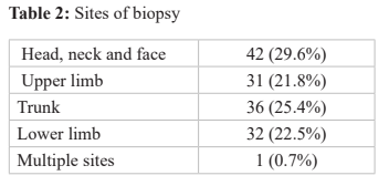
performed. Head, neck and face were the commonest
sites of biopsies (29.6%). The most common
biopsy technique was incisional type in 70 (50.4%).
Histopathology showed granulomatous features in 49
(34.8%) cases, out of which tuberculoid type was the
commonest, in 29 (58.0%). Positive clinicopathological
correlation was seen in 117/142 (82.4%) for all biopsies
and 41/49 (85%) for granulomaous skin lesions.
Conclusion: Tuberculoid type was the
most common cutaneous granuloma. High
clinicopathological correlation in our study supports
histopathology as an important tool for diagnosis
of granulomatous as well overall skin disorders.
References
M, Sathian B. Profile of skin biopsies and patterns
of skin cancer in a tertiary care center of Western
Nepal. Asian Pacific Journal of Cancer Prevention.
2015;16(8):3403-3406.
2.Gautam, K., Pai, R., & Bhat, S.. Granulomatous
lesions of the skin. Journal of Pathology of
Nepal, 2011;1(2), 81-86.
3. Weedon D. The granulomatous reaction patterns.
In: Weedon’s Skin Pathology. 3rd ed: Churchill
Livingstone Elsevier; 2010. 170-194.
4.Chakrabarti S, Pal S, Biswas BK, Bose K, Pal S,
Pathak S. Clinico-pathological study of cutaneous
granulomatous lesions-a 5 yr experience in a tertiary
care hospital in India. Iranian journal of pathology.
2016;11(1):54.
5. Elston DM, Stratman EJ, Miller SJ. Skin biopsy:
biopsy issues in specific diseases. Journal of the
American Academy of Dermatology. 2016 Jan
;74(1):1-6.
6. Stewart MI, Bernhard JD, Cropley TG, Fitzpatrick
TB. The structure of skin lesion and fundamentals
of diagnosis. In: Freedberg IM, Eisen AZ, Wolff
K, Austen KF, Goldsmith LA, Katz SI, editors.
Fitzpatrick’s Dermatology in general medicine. 6th
ed. McGraw-Hill: New York; 2003. 11-30
7.Korfitis C, Gregoriou S, Antoniou C, Katsambas
AD, Rigopoulos D. Skin biopsy in the context of
dermatological diagnosis: a retrospective cohort
study. Dermatology research and practice. 2014 Jan
1;2014.
8. Shrestha DP, Gurung D, Rosdahl I. Prevalence of
skin diseases and impact on quality of life in hilly
region of Nepal. Institute of Medicine. Journal.
2012;34(3):44-49.
9. Carli P, de Giorgi V, Chiarugi A, Nardini P,
Weinstock MA, Crocetti E, Stante M, Giannotti B.
Addition of dermoscopy to conventional naked-eye
examination in melanoma screening: a randomized
study. Journal of the American Academy of
Dermatology. 2004 May ;50(5):683-689.
10. Zafar MN, Sadiq S, Memon MA. Morphological
study of different granulomatous lesions of the skin.
Journal of Pakistan Association of Dermatology.
2016 Dec ;18(1):21-28.
11. Dhar S, Dhar S. Histopathological features of
granulomatous skin diseases: an analysis of 22 skin
biopsies. Indian Journal of Dermatology. 2002 Apr
;47(2):88.
12. Rajbhandari A, Adhikari RC, Shrivastav S,
Parajuli S. Histopathological study of cutaneous
granulomas. Journal of Pathology of Nepal. 2019
Sep ;9(2):1535-1541.
13. Adhikari RC, Shrestha KB, Sayami G.
Granulomatous inflammation: a histopathological
study. Journal of Pathology of Nepal. 2013 Oct
;3(6):464-468.
14. Pawale J, Belagatti SL, Naidu V, Kulkarni MH.
Histopathological study of cutaneous granuloma.
Indian Journal of Public Health Research &
Development. 2011;2(2):74-79.
15.Bal A, Mohan H, Dhami GP. Infectious
granulomatous dermatitis: A clinico pathological
study. Indian journal of dermatology. 2006 Jul
;51(3):217.
16. Bansal C, Batra M, Sharma KL, Tulsyan S,
Srivastava AN. Facial granulomatous dermatoses:
A clinico-pathological study. Journal of the Saudi
Society of Dermatology & Dermatologic Surgery.
2013 Jul ;17(2):55-61.
17. El-Khalawany M, Meraag I, Eassa B, El-Naby
HH. Clinicopathological features and the practice
of diagnosing infectious cutaneous granulomas in
Egypt. International Journal of Infectious Diseases.
2011 Sep ;15(9):e 620-626.
18. Qureshi R, Sheikh RA, ul Haque A. Chronic
granulomatous inflammatory disorders of skin at
a tertiary care hospital in Islamabad. International
Journal of Pathology. 2018 Oct 23:31-34.
19. Gupta K, Kumari A, Mangal K.
Granulomatous lesions: a diagnostic challenge to
dermatopathologists. Int J Med Res Professionals.
2016;2(4):33-39.
20. Kafle SU, Chaudhary D, Yamu SY, Jha K.
Diagnostic Significance of Clinicopathological
Concordance in Various Spectrums of Skin
Disorders. Birat Journal of Health Sciences. 2020
Jun 26;5(1):955-959.
21. Makwana V, Patel N, Shah A, Makwana S.
Granuloma Revisited: a Prospective Study of
Granulomatous Skin Lesions at a Tertiary Care
Centre In Gujarat. International Journal of Recent
Scientific Research. 2018Aug;9(8):28607-28613.